Immunofluorescence staining of C2BBe1 intestinal epithelial cells infected with Y. pseudotuberculosis.
Dr. Michael Böhringer, FLI, Germany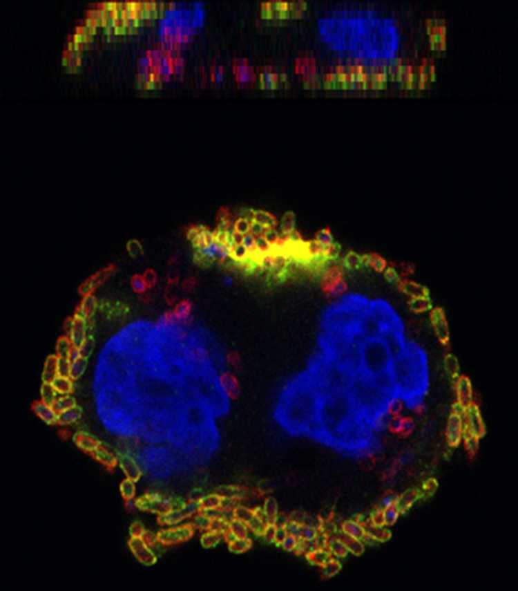
Yersinia pseudotuberculosis is a Gram-negative bacillus that causes self-limiting colitis, ileitis, or mesenteric lymphadenitis with possible pseudoappendicitis symptoms in humans. Reactive arthritis can appear in the aftermath, and, occasionally, the septic-typhoid disease occurs. The primary sources of human infection are contaminated water or food, especially pork products, and direct transmission from animals. Y. pseudotuberculosis is distributed worldwide, especially in the temperate and subtropical climate zone. Animal reservoirs are rodents, lagomorphs, and wild birds, but infections can also affect farm, zoo, and companion animals, either inapparent or presenting as septicaemia or pseudotuberculosis. For example, Y. pseudotuberculosis is detected regularly in the faeces of clinically inapparent dogs, but fatal outbreaks in zoo animals also occur. In Germany, Y. pseudotuberculosis infections are most frequently caused by strains of the serogroup O:1, and occasionally by strains of the groups O:2 and O:3; however, all Y. pseudotuberculosis strains must be considered as pathogenic.
Methodical long: C2BBe1 cells grown on glass coverslips were incubated with Y. pseudotuberculosis for two hours. After infection, samples were fixed with formaldehyde. For differential staining of intra- and extracellular bacteria, samples were first incubated with polyclonal anti-Y. pseudotuberculosis antibody (Abcam, ab26120) combined with FITC-labelled secondary antibody, followed by permeabilization with Triton X-100 and subsequent incubation with anti-Y. pseudotuberculosis antibody and CF647-labelled secondary antibody. Cell nuclei were stained with DAPI. Images were recorded with a confocal laser-scanning microscope. The central picture shows one layer of an image stack with corresponding images of the z-axis on the right and above. Red color indicates intracellular bacteria, and extracellular bacteria appear yellow, DAPI staining is coloured blue.
Methodical short: Image of C2BBe1 intestinal epithelial cells infected with Y. pseudotuberculosis in vertical and side views. Differential staining shows intracellular bacteria in red. Extracellular bacteria appear yellow, and cell nuclei are coloured blue.